38 onion cells under microscope with labels
Onion Cells Under a Microscope (100x-2500x) - YouTube In this video you will see onion cells under a microscope (100x-2500x) as is, without any coloring. To observe the onion cells the thin membrane is used. It... Onion Cells Microscope Stock Photos and Images - Alamy Onion cells under the microscope. Garden onion, Bulb Onion, Common Onion (Allium cepa), cell tissue of a garden onion with dyed chromosomes, light microscopy, x 120, Germany. Onion Cells under the Microscope. Onion skin cells under the microscope, horizontal field of view is about 0.61 mm. Detailed view of the cells of a red onion as seen ...
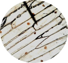
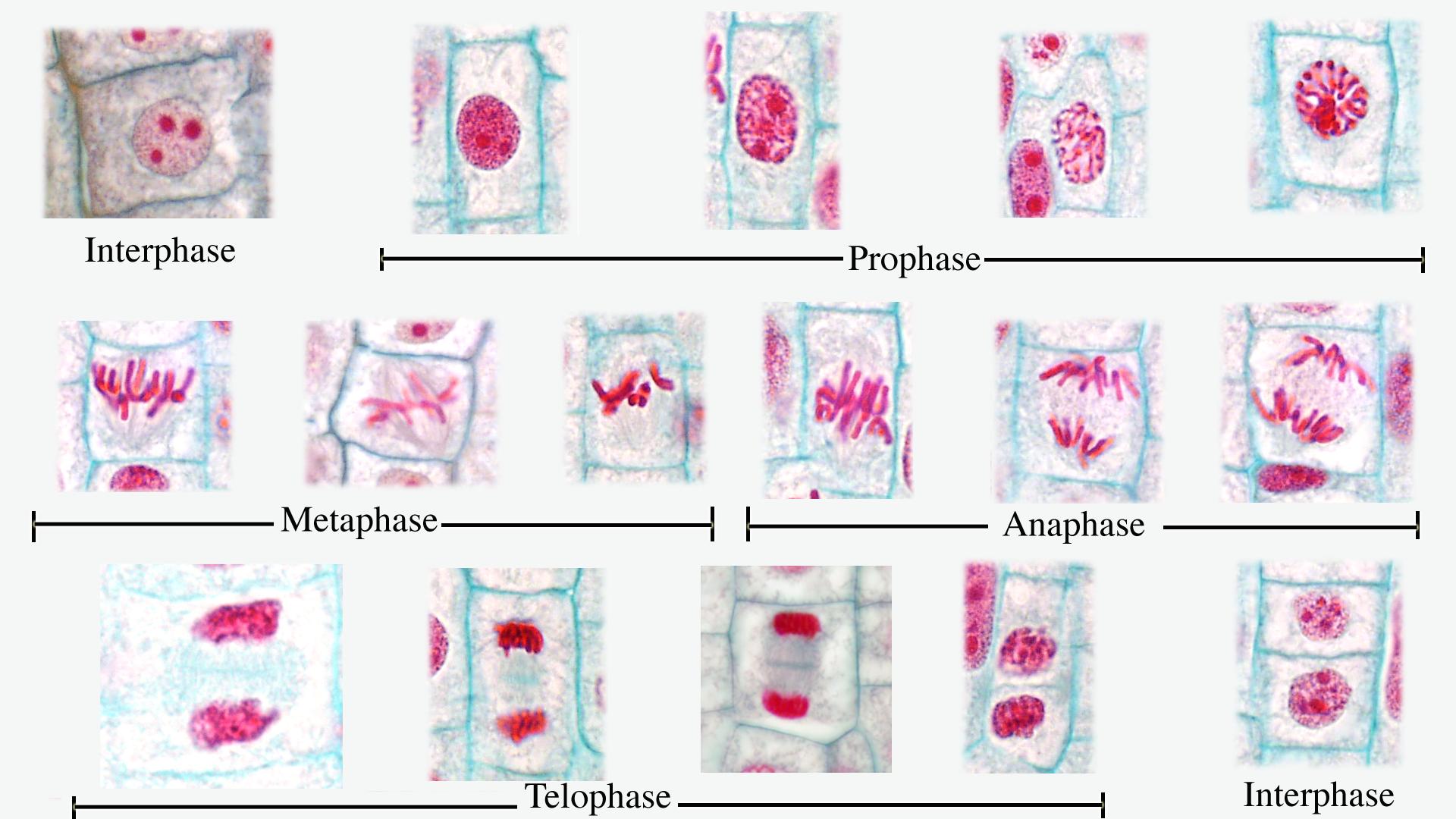
Plant tissue under a microscope - xylem and phloem - Rs' Science The highly active mitosis area is highlighted with a red dash line. Within that area, you can easily find cells undergoing different phases of mitosis, prophase , metaphase , anaphase, and telophase. (Modified from the guidebook of Rs' Science - 25 Microscope Prepared Slide Set) The Stem - Xylem and Phloem

Onion cells under microscope with labels
Onion Cell Lab Report.docx - Onion Cell Lab Report By Onion Cell Lab Report By : Nawaf Almalki Introduction: Many things that are viewed using a microscope, particularly cells, can appear quite transparent under the microscope. The internal parts of the cells, the organelles, are so transparent that they are often difficult to see. Biologists have developed a number of stains that help them see the cells and their organelles by adding color to ... Microscopy, size and magnification - Microscopy, size and ... - BBC Place cells on a microscope slide. Add a drop of water or iodine (a chemical stain). Lower a coverslip onto the onion cells using forceps or a mounted needle. This needs to be done gently to... The following diagram shows cells of onion peel label class ... - Vedantu In order to label them, we need to understand its anatomy and know about various structures present in it. Onion peel is an example of a plant cell whereas a human cheek cell is an example of an animal cell. Complete answer:
Onion cells under microscope with labels. Onion Cell Under Labeled Microscope draw and label!) Remove the slide from the microscope Start with the low power objective and work your way until you have focused the Onion cell using the medium power objective Then place the onion skin onto the center of the slide A scientist is observing onion cells and human cheek cells under a microscope Add 2 drops of iodine (or other stain) to the onion slide Add 2 drops of iodine (or ... Onion cell Images, Stock Photos & Vectors - Shutterstock Find Onion cell stock images in HD and millions of other royalty-free stock photos, illustrations and vectors in the Shutterstock collection. Thousands of new, high-quality pictures added every day. Plant Cell Under Microscope Labeled 40X : Young Root 2 Of Broad Bean ... Cells and viewing them under the microscope. A small square of a red onion skin (membrane) was observed under a microscope at high power (x40) magnification. (iv) describe how you applied the stain. They must draw and label the nucleus, cell membrane set up your microscope, place the onion root slide on the stage and focus on low (40x) power. Onion Skin Cells - Investigation - Exploring Nature 5. Observe the onion tissue under the microscope at 4x, 10x and 40x with lots of light (open diaphragm). Then slowly close the diaphragm while observing the image to find the best light for seeing cellular details. 6. Draw a section of onion skin cells at 10x magnification. Then switch to 40x and draw one cell and label it.
DOC Plant and Animal Cells Microscope Lab - Hillsboro City Schools Make a drawing of one onion cell, labeling all of its parts as you observe them. (At minimum you should observe the nucleus, cell wall, and cytoplasm.) Cheek cells 1. To view cheek cells, gently scrape the inside lining of your cheek with a toothpick. DO NOT GOUGE THE INSIDE OF YOUR CHEEK! (We will observe blood cells in a future lab!!) 2. How to observe onion cells under a microscope? - JacAnswers How to observe onion cells under a microscope? Gently lay a microscopic cover slip on the membrane and press it down gently using a needle to remove air bubbles. Touch a blotting paper on one side of the slide to drain excess iodine/water solution, Place the slide on the microscope stage under low power to observe. Plant Cell Under Microscope 40X Labeled : 1 - Chloroplast and cell wall ... The different images below were taken with two different types of microscopes. 1.can only turn fine adjustment 2.draw one row of cells across the middle 3.label the chloroplasts and cell wall. When using the microscope always start by focusing under low power and working your way up to high power. Onion Skin Cells Labeled - the wonderful microworld onion skin cells ... Here are a number of highest rated Onion Skin Cells Labeled pictures on internet. We identified it from reliable source. Its submitted by direction in the best field. We resign yourself to this nice of Onion Skin Cells Labeled graphic could possibly be the most trending topic in the same way as we allowance it in google help or facebook.
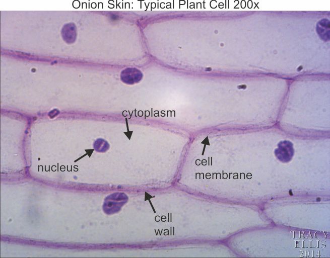
Onion Skin Cells Labeled - the wonderful microworld onion skin cells ... Onion Skin Cells Labeled. Here are a number of highest rated Onion Skin Cells Labeled pictures on internet. We identified it from obedient source. Its submitted by executive in the best field. We... DOC The Onion Cell Lab - chsd.us Onion tissue provides excellent cells to study under the microscope. The main cell structures are easy to see when viewed with the microscope at medium power. For example, you will observe a large circular . nucleus. in each cell, which contains the genetic material for the cell. In each nucleus, are round bodies called . nucleoli Oxford Cambridge and RSA Friday 16 October 2020 – Morning 1 (a) A student was observing onion epithelial cells using a light microscope. They photographed these cells and the image obtained is shown in Fig. 1.1. The student then made a drawing of a few cells from this image. The drawing is shown in Fig. 1.2. Fig. 1.1 cytoplasm cell wall large permanent vacuole ribosome Fig. 1.2 Onion Epidermis - kuensting.org Onion epidermal cells, iodine stain, 400X. The nucleus of an onion epidermal cell, 1000X magnification. ...
swifty science: onion cell lab
Onion Microscope Under Cell Labeled Phases of the cell cycle Label the cell wall, cytoplasm, and the pigmented organelle structures (but you must label it with their real name When observing an onion cell under the microscope, it appear to be long an oval in shape move your slide so that your field of view is centered on the root tip Label structures within the cell Bobby Rush ...

Onion Cell Under Microscope Mitosis - Micropedia
Observing Onion Cells Under The Microscope Afterwards, carefully mount the prepared and stained onion cell slide onto the microscope stage. Make sure that the cover slip is perfectly aligned with the microscope slide, and that any excess stain has been wiped off. Secure the slide on the stage using the stage clips.
Cell Biology
What organelles are in an onion cell? - Biology Stack Exchange To answer your question, onion cells (you usually use epithelial cells for this experiment) are 'normal' cells with all of the 'normal' organelles: nucleus, cytoplasm, cell wall and membrane, mitochondria, ribosomes, rough and smooth endoplasmic reticulum, centrioles, Golgi body and vacuoles.

Fanos' MCB Blog: Onion Skin
Cells and Reproduction - BBC Bitesize Onion cells are easy to see using a light microscope. ... A small tube placed under the skin of the upper arm. ... Five small tubes with labels and stoppers or lids Cress seeds Labels Cotton wool ...
Microscope 400x Cheek Cells Under A Microscope - Micropedia
Under the Micrsocope: Onion Cell (100x - 400x) - YouTube In this "experiment" we will see onion cells under the microscope.For the experiment you will only need onion, dropper and the microscope (container and tool...

scyhighbio: microscope lab
Animal Cell Diagram Under Microscope Labeled - ACTUINDE Animal Cell Diagram Under Microscope Labeled. Animal Cell Diagram Under Microscope. Function cell does in the body dictate the change and adaptation done by cell. When observing onion cells, there is the Cell Surface Membrane which is present in all living cells. We all keep in mind that the human body is quite intricate and a method I ...
Composite of all stages of mitosis in onion root tip - labeled - UWDC - UW-Madison Libraries
Microscope Onion Cell Diagram - Wiring Schematic Online Sketch the onion peel cell as seen under the microscope label the. The cheek epithelium cell is the only one that has centrioles the barrel shaped organelle that is responsible for helping organize chromosomes during cell division. Preparation and viewing of onion cells expected time for completion. Observe an onion cell under the microscope.
Onion skin 200x « Dissection Connection
Epidermal onion cells under a microscope. Plant cells appear polygonal ... Observing onion cells under the microscope. For this microscope experiment, the thin membrane will be used to observe the cells. An easy beginner experiment. Jessica Williams. Ideas for Work. Similar ideas popular now. Applied Science. Subjects. Physical Science. Technology.
Post a Comment for "38 onion cells under microscope with labels"