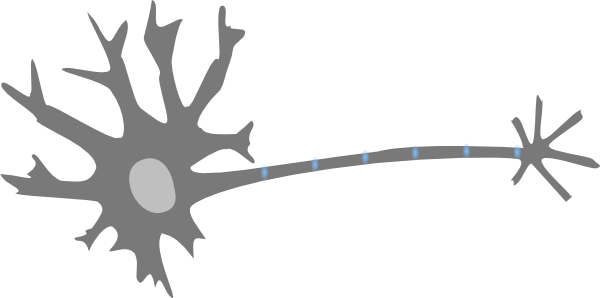
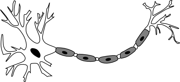
42 nerve cell with labels
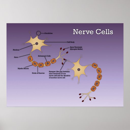
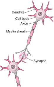
Histology of neurons: Morphology and types of neurons - Kenhub A neuron (nerve cell) is a specialized cell that conveys electrochemical impulses throughout the body. The cytology of a neuron facilitates the transmission of either: 'top-down' information from the brain to the periphery, via efferent neurons (e.g. to permit locomotion) (efferent neurons) or, Nerve: anatomy, definition, types, functions | Kenhub An individual nerve cell (neuron) is made up of small branching extensions called dendrites, a cell body (soma), and an axon which is one single, long branch. At the end of the axon, we find the axon terminals. Axon terminals meet the dendrites of adjoining neurons at the synaptic cleft.
Nerve - Plexus Worldwide Nerve is a specially formulated combination of vitamins, minerals, herbs, and amino acids to help support healthy nerve cells and nervous system. Don't let nerve discomfort get in the way of having a good day every day - get Plexus Nerve.* ... Nutrition Label: Recommended Servings: Adults take 1 capsule twice daily with or without food ...

Nerve cell with labels
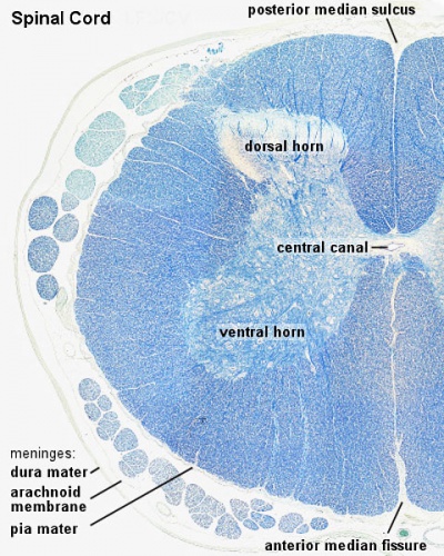
Neuron Labeling Worksheet - Nervous System Label The Neuron Label the parts of the neuron with the correct title: Myelin sheath nodes of ranvier. Draw a picture of a spinal cord. Click on the template below to see it in its own window and . Label the central canal, grey matter, white matter, spinal nerve. Consider this main learning worksheet to teach all about the fabulous neuron cells and the 6 parts. Cells of the Nervous System - Neurons - TeachMePhysiology The nervous system comprises of two groups of cells, glial cells and neurones. Neurones are responsible for sensing change in their environment and communicating with other neurones via electrochemical signals. Glial cells work to support, nourish, insulate neurones and remove the waste products of metabolism. Myelin Sheath Function & Type of Conduction | Schwann Cells vs ... The nerve cells in your body have a protrusion extending out of the soma that we call an axon. If your smartphone is the soma, then the axon sort of looks like the cord coming out of the ...
Nerve cell with labels. Nerve Cells (Neurones) and Synapses Diagram Worksheets NERVE CELLS AND SYNAPSES DIAGRAM WORKSHEET Included in this resource: Nerve Cell Diagram Worksheet - Looking at parts of the nerve cell, students can label and describe the functions (e.g. nucleus, axon, dendrites, myelin sheath etc). Students are also asked to define what a neuron is and the three types or neurone, linked to a simple diagram. Glial Cells | Location, Structure, Summary & Function Summary: Glial cells are essentially any of several kinds of cells that principally focus on supporting nerve cells. Glial cells are found in the central as well as the peripheral nervous system, alongside nerve cells. Glial cells have a fibrous appearance due to thick bundles of cytoplasmic filaments. The five types of glial cells found in the ... correctly label the following anatomical features of a nerve The nerve is a tiny bundle of neurons that travels within the body and is responsible for communicating between the brain and the rest of the body. As the brain ages, the nerve becomes stretched and distorted. The nerve is also the target of many forms of disease like multiple sclerosis, Parkinson's disease, and spinal cord injury. Neuromuscular Junction Structure and Functions - New Health Advisor Acetylcholine is the neurotransmitter secreted by the somatic motor neurons. There are receptors of acetylcholine present in the skeletal muscle cells. So this secreted acetylcholine then passes the cleft by diffusion and bind with the receptors. They are like puzzle pieces which fit or key which opens the door.
Neurons: Meaning, Types, Functions, Diagrams - Embibe Cell body or Soma: The cell body or Soma is also called a cyton. It contains a nucleus and cytoplasm that connects to dendrites. It carries signals to the other neurons and controls all the functions of the cell. Axon: Axon is a part of a nerve cell or neuron that carries nerve impulses away from the cell body. A neuron typically has one long ... Spinal Nerves: Anatomy, Function, and Treatment - Verywell Health There are 31 pairs of spinal nerves: 2 Eight cervical spinal nerves on each side of the spine called C1 through C8 Twelve thoracic spinal nerves in each side of the body called T1 through T12 Five lumbar spinal nerves on each side called L1 through L5 Five sacral spinal nerves in each side called S1 through S5 Synapses in the Nervous System - Verywell Health What Synapses Do Parts of the Synapse Types In the central nervous system, a synapse is a small gap at the end of a neuron that allows a signal to pass from one neuron to the next. Synapses are found where nerve cells connect with other nerve cells. Synapses are key to the brain's function, especially when it comes to memory. 1 Neuroanatomy, Visual Pathway - StatPearls - NCBI Bookshelf The ganglion cell layer and nerve fiber layer serve as the foundation of the optic nerve; the former contains the cell bodies, and the latter contains the axons as they stream across the retina. It consists of two types of fibers, namely temporal and nasal fibers, which control the nasal and temporal parts of the visual field, respectively. ...
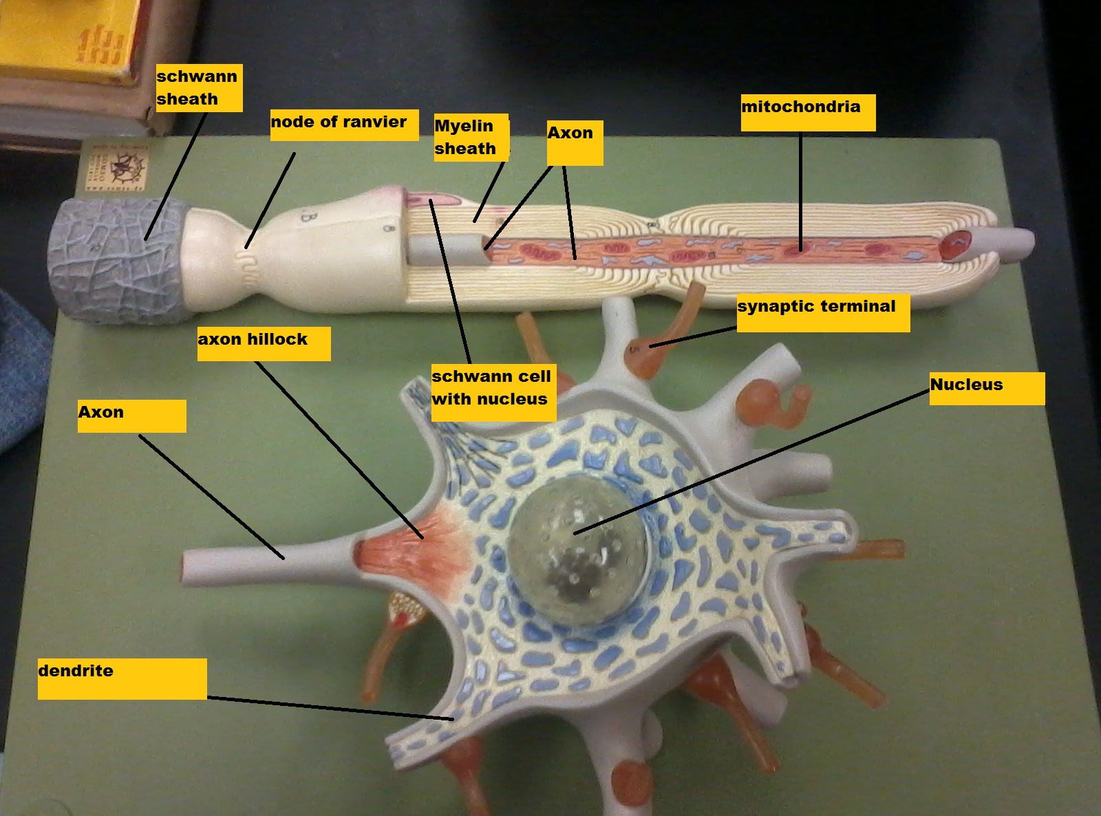
CBSE Class 9 Science Practicals Identification of parenchyma ... 5) Fibers and sclerites are two types of parenchyma cells. B) Aim: To identify striped, smooth and cardiac muscle fibers and nerve cells in animals, from prepared slides. Draw their labeled diagrams. Requirements: Prepared slides of striped, smooth and cardiac muscle fibers and nerve cells in animals and compound microscope. Procedure: These Are the 12 Cranial Nerves and Their Functions - Healthline The optic nerve is the sensory nerve that involves vision. When light enters your eye, it comes into contact with special receptors in your retina called rods and cones. Rods are found in large... Free Nervous System Worksheets and Printables The Nervous System Worksheets and Brain Game - These worksheets and games introduce kids to concepts such as nerve cells and electrical impulses. Cranial Nerves and Brain Coloring Page - This brain coloring page teaches kids about the cranial nerves. The Brain Labeling Sheet - This is a fun and easy brain labeling sheet for younger kids. Neuron under Microscope with Labeled Diagram - AnatomyLearner But, first, let's try to identify the following features from a neuron with the help of a labelled diagram. Cell body or perikaryon of a neuron Nucleus, cytoplasm, the plasma membrane of a neuron Nissl bodies in the cell body of a neuron An initial segment of axon and axon hillock Dendrites and axons of a neuron Axolemma and myelin sheath

Answer: Ventral Horn (grey matter).
What Are Nerve Cells? - Function, Types & Structure Bipolar neurons are nerve cells that have two projections and are used in specialized sense detection, such as in smell and sight. Multipolar neurons are nerve cells that have many cell extensions,...

Make a Sketch of the Human Nerve Cell. What Function Do Nerve Cells Perform? - Science | Shaalaa.com
Nervous System of Earthworm - The Biology Notes 2. Peripheral nervous system. From the brain 8 to 10 pairs of nerves arises which supply to the prostomium, buccal cavity, and pharynx. 2 pairs of nerves arise from the circumpharyngeal connectives supply 1 st segment and buccal cavity.; From the sub-pharyngeal ganglia, 3 pairs of the nerve arise supply to 2 nd, 3 rd, and 4 th segments.; Each segmental ganglion of the ventral nerve cord gives ...

Rens blog : Science, cells
Schwann Cell Anatomy - Human Anatomy - GUWS Medical Figure 25.1 Label this diagram of a motor neuron. Figure 25.2 Label the features of the myelinated nerve fiber. Figure 25.3 Micrograph of a multipolar neuron and neuroglia from a spinal cord smear (100x micrograph enlarged to 600x). -Nerve fiber (axon) general name for processes (either dendrites or axon) of the neuron.

Histology Drawings: February 2014
Visual Guide to Your Nervous System - WebMD Your Command Central. Made up of billions of nerve cells called neurons, your nervous system is what lets you do everything from breathe to walk to dream. It has two main parts: the central ...
ANAT2511 Introduction to Histology - Embryology
Draw And Label Diagram Of Animal Cell : Draw A Neat Labelled Diagram Of ... The first is a colored and labeled cell diagram. Source: pixfeeds.com. Draw an animal cell and label it. Source: . The animal cell is made up of several structural organelles enclosed in the plasma membrane, that enable it to function properly, eliciting mechanisms that benefit the host (animal). Source:

Nerve Cells |authorSTREAM
Lab 4: Nervous System - Biology LibreTexts Introduction: In this lab, we will explore the anatomy & physiology of the nervous system. Nervous systems are unique to animals and are critical for detecting and interpreting information, making decisions, and regulating body functions and movements. Nervous systems are constructed from neurons and glia. Neurons are the main functional cells ...
Nerve Cells Diagram - ClipArt Best
Neuroanatomy, Unmyelinated Nerve Fibers - NCBI Bookshelf Myelination is a crucial positive factor in determining the regeneration and repair of injured nerve fibers. It enables the afferent axons to regenerate into the peripheral nerve stump along the Schwann cell tubes. On the other hand, unmyelinated axons have lesser regenerative potential, even with intact Schwann cell tubes, such as nerve crush.
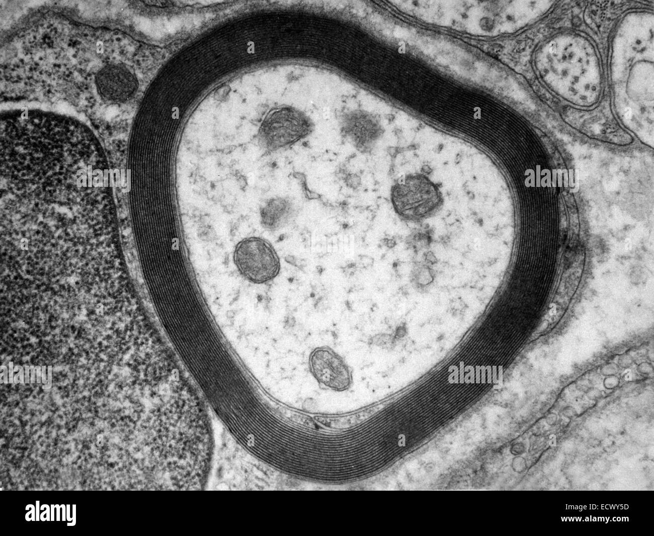
transmission electron micrograph of myelin sheath Stock Photo: 76787465 - Alamy
What are the 12 cranial nerves? Functions and diagram Scientists use Roman numerals from I to XII to label the cranial nerves in the brain. The 12 cranial nerves include the: olfactory nerve optic nerve oculomotor nerve trochlear nerve trigeminal...
Nerve Cells Diagram Posters | Zazzle
Synaptic Cleft | Anatomy, Structure, Diseases & Functions A synaptic cleft is a space that separates two neurons. It forms a junction between two or more neurons and helps nerve impulse pass from one neuron to the other. In this article, we will talk about different aspects of synaptic cleft, its anatomy, and functions. You will completely understand the concept of synapse after reading this article.
Dead Neuron Clip Art at Clker.com - vector clip art online, royalty free & public domain
Neuronal Cell Markers The protein is detectable in both embryonic and adult neurons with the exception of cerebellar Purkinje cells, olfactory bulb mitral cells, retinal photoreceptor cells, and dopaminergic neurons in the substantia nigra [ 33, 34 ]. [enlarge] Figure 2. Example of NeuN-labeled neurons in mouse neocortex. Reproduced from Figure 3B of [ 2 ].

The Nerve Cell
What Is a Neuron? Diagrams, Types, Function, and More - Healthline Nervous system cells are called neurons. They have three distinct parts, including a cell body, axon, and dendrites. These parts help them to send and receive chemical and electrical signals. While...
Chapter 14 Nervous Tissue - Biology 4 Human AnatomyProfessor Julie GallagherBarstow Community ...
Myelin Sheath Function & Type of Conduction | Schwann Cells vs ... The nerve cells in your body have a protrusion extending out of the soma that we call an axon. If your smartphone is the soma, then the axon sort of looks like the cord coming out of the ...

Science: NERVOUS SYSTEM
Cells of the Nervous System - Neurons - TeachMePhysiology The nervous system comprises of two groups of cells, glial cells and neurones. Neurones are responsible for sensing change in their environment and communicating with other neurones via electrochemical signals. Glial cells work to support, nourish, insulate neurones and remove the waste products of metabolism.

Nerve Cells Educational Resources K12 Learning, Life Science, Science Lesson Plans, Activities ...
Neuron Labeling Worksheet - Nervous System Label The Neuron Label the parts of the neuron with the correct title: Myelin sheath nodes of ranvier. Draw a picture of a spinal cord. Click on the template below to see it in its own window and . Label the central canal, grey matter, white matter, spinal nerve. Consider this main learning worksheet to teach all about the fabulous neuron cells and the 6 parts.
Why don't nerve cells repair? - Quora
What is another name for a nerve cell? - Quora
Neuron B&w Clip Art at Clker.com - vector clip art online, royalty free & public domain
Overview of the Peripheral Nervous System - Brain, Spinal Cord, and Nerve Disorders - Medicine.com
Post a Comment for "42 nerve cell with labels"