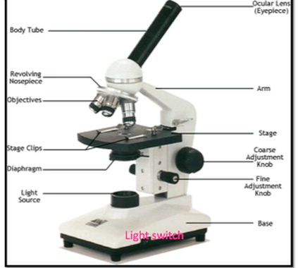
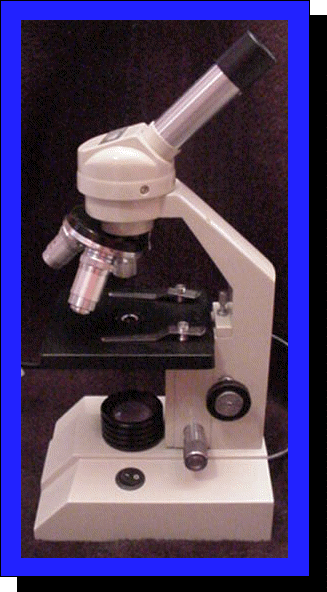
43 light microscope with labels
Light Labs distributes PCR... Welcome to Light Labs. Since 2002, Light Labs has distributed high quality laboratory consumables and equipment, including MultiMax Barrier tips, PCR tubes and strip tubes, PCR plates, and much more. With an emphasis on customer service, we have successfully served the research marketplace with a wide array of laboratory goods. An Introduction to the Light Microscope, Light Microscopy Techniques ... For example, using green light with a wavelength of 550 nm and an objective with a typical NA of 0.7, a standard light microscope can resolve features down to a limit of 0.61 × ... Examples include biological samples that are intrinsically fluorescent or have been labeled with a fluorescent marker, as well as single molecules and other ...
umassmed.edu › cemf › whatisemWhat is Electron Microscopy? - UMASS Medical School Conventional scanning electron microscopy depends on the emission of secondary electrons from the surface of a specimen. Because of its great depth of focus, a scanning electron microscope is the EM analog of a stereo light microscope. It provides detailed images of the surfaces of cells and whole organisms that are not possible by TEM.

Light microscope with labels
Parts of the Microscope with Labeling (also Free Printouts) 5. Knobs (fine and coarse) By adjusting the knob, you can adjust the focus of the microscope. The majority of the microscope models today have the knobs mounted on the same part of the device. Image 5: The circled parts of the microscope are the fine and coarse adjustment knobs. Picture Source: bp.blogspot.com. Light Microscopes - an overview | ScienceDirect Topics J.H. Holgate, J. Webb, in Encyclopedia of Food Sciences and Nutrition (Second Edition), 2003 Principles. The light microscope is an instrument for visualizing fine detail of an object. It does this by creating a magnified image through the use of a series of glass lenses, which first focus a beam of light onto or through an object, and convex objective lenses to enlarge the image formed. Parts of Stereo Microscope (Dissecting microscope) - labeled diagram ... Labeled part diagram of a stereo microscope Major structural parts of a stereo microscope. There are three major structural parts of a stereo microscope. The viewing Head includes the upper part of the microscope, which houses the most critical optical components, including the eyepiece, objective lens, and light source of the microscope.
Light microscope with labels. ProSciTech Laboratory supplies and Lab equipment for Histology, Pathology, Light Microscopy, Electron Microscopy and specialist researchers. en.wikipedia.org › wiki › FluorescenceFluorescence - Wikipedia Fluorescence is the emission of light by a substance that has absorbed light or other electromagnetic radiation.It is a form of luminescence.In most cases, the emitted light has a longer wavelength, and therefore a lower photon energy, than the absorbed radiation. Label a Microscope - Storyboard That In this activity, students will create a poster of a microscope with labeled parts. Students will identify and describe the microscope parts and functions. This is an awesome activity to complete at the beginning of either the school year or the unit on basic cells. ... Provides light to illuminate the specimen, sometimes a mirror is also used ... Compound Light Microscopes | Products | Leica Microsystems Compound light microscopes from Leica Microsystems know what counts: superlative image quality, ergonomic handling, fast results. Products. Light Microscopes ... Microscope Cameras. Microscope Software. Aivia Free Trial. Microscope Objective Lens. Objectivefinder Objective Classes Labeling of Objectives Leitz Optic Center.
Required practical - using a light microscope - BBC Bitesize To use a light microscope to examine animal or plant cells. To make observations and draw scale diagrams of cells. Method. Rotate. Rotate the objective lenses so that the low power, eg ×10, is in ... Light Microscope- Definition, Principle, Types, Parts, Labeled Diagram ... A light microscope is a biology laboratory instrument or tool, that uses visible light to detect and magnify very small objects and enlarge them. They use lenses to focus light on the specimen, magnifying it thus producing an image. The specimen is normally placed close to the microscopic lens. › seterra › en-anThe Eye - Science Quiz - Seterra The Eye - Science Quiz: Our eyes are highly specialized organs that take in the light reflected off our surroundings and transform it into electrical impulses to send to the brain. The anatomy of the eye is fascinating, and this quiz game will help you memorize the 12 parts of the eye with ease. Light enters our eyes through the pupil, then passes through a lens and the fluid-filled vitreous ... Student's Guide: How to Use a Light Microscope An ultraviolet microscope uses UV light to view specimens at a resolution that isn't possible with the common brightfield microscope. It utilizes UV optics, light sources, as well as cameras. Because of the shorter wavelengths of UV light (180-400 nm), the image produced is clearer and more distinct at a magnification approximately double what ...
Microscope Objective Lens | Products | Leica Microsystems The objective lens is a critical part of the microscope optics. The microscope objective is positioned near the sample, specimen, or object being observed. It has a very important role in imaging, as it forms the first magnified image of the sample. The numerical aperture (NA) of the objective indicates its ability to gather light and largely determines the microscope’s … Light Microscope - an overview | ScienceDirect Topics 6.5 Microscopy. The light microscope is an important tool in the study of microorganisms, particularly for identification purposes. The compound light microscope uses visible light to directly illuminate specimens in a two-lens system, resulting in the illuminated specimen appearing dark against a bright background. Label the light microscope | Teaching Resources Label the light microscope. Subject: Biology. Age range: 11-14. Resource type: Worksheet/Activity (no rating) 0 reviews. Science Resources. 4 1 reviews. ... Low ability worksheet, label the main parts of a microscope, key terms given at the bottom of the worksheet. Creative Commons "Attribution" Compound Microscope: Definition, Diagram, Parts, Uses, Working ... - BYJUS Compound microscope is a type of optical microscope that is used for obtaining a high-resolution image. There are more than two lenses in a compound microscope. Learn about the working principle, parts and uses of a compound microscope along with a labeled diagram here.
Types Of Light Microscopes
› microscopy › intZEISS Axioscan 7 Microscope Slide Scanner Digitize your specimens with Axioscan 7 – the reliable, reproducible way to create high-quality virtual microscope slides. Axioscan 7 combines qualities that you would not expect to get in a slide scanner: high speed digitization and outstanding image quality plus an unrivaled variety of imaging modes are all available in a fully automated and easy to operate system.
Using a Light Microscope - AyushiSinhaMicroscopy
Electron microscope - Wikipedia An electron microscope is a microscope that uses a beam of accelerated electrons as a source of illumination. As the wavelength of an electron can be up to 100,000 times shorter than that of visible light photons, electron microscopes have a higher resolving power than light microscopes and can reveal the structure of smaller objects.. Electron microscopes use shaped magnetic …

Euglena Acus 2 - BF microscope 1250x - YouTube
Microscope Drawing And Label - Painting Valley All the best Microscope Drawing And Label 33+ collected on this page. Feel free to explore, study and enjoy paintings with PaintingValley.com. ... Compound Light Microscope Drawing. Simple Microscope Drawing. Microscope Drawing. Microscope Drawing Circles. Philippine Map Drawing With Label.
Using the Microscope
Microscope labels Flashcards | Quizlet Microscope label Learn with flashcards, games, and more — for free.

Stratified squamous non-keratinized epithelium
Labeling the Parts of the Microscope Labeling the Parts of the Microscope. This activity has been designed for use in homes and schools. Each microscope layout (both blank and the version with answers) are available as PDF downloads. You can view a more in-depth review of each part of the microscope here.

Unlabeled Compound Light Microscope Diagram - Micropedia
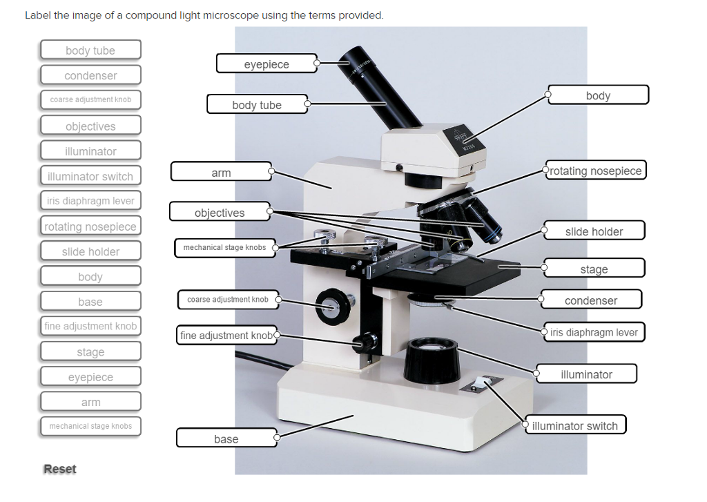
Compound Microscope Parts - Labeled Diagram and their Functions - Rs ... There are three major structural parts of a compound microscope. The head includes the upper part of the microscope, which houses the most critical optical components, and the eyepiece tube of the microscope. The base acts as the foundation of microscopes and houses the illuminator. The arm connects between the base and the head parts.

Trypanosoma | Medical Laboratories
Simple Microscope - Diagram (Parts labelled), Principle, Formula and Uses It is a type of optical microscope that uses visible light and lens to magnify objects. Despite the fact that they are rudimentary imaging devices, simple microscope finds use in microbiology to study biological specimens and microscopic organisms such as fungi, hydra and algae. They are also used by pedologists to study soil samples and ...
Solved: Label The Image Of A Compound Light Microscope Usi... | Chegg.com
Parts of a microscope with functions and labeled diagram Q. Differentiate between a condenser and an Abbe condenser. Ans. Condensers are lenses that are used to collect and focus light from the illuminator into the specimen. They are found under the stage next to the diaphragm of the microscope. They play a major role in ensuring clear sharp images are produced with a high magnification of 400X and above.

Microscope With Labels vector, free vector graphics - Vector.me
Microscope Labeling - The Biology Corner Microscope Labeling. This simple worksheet pairs with a lesson on the light microscope, where beginning biology students learn the parts of the light microscope and the steps needed to focus a slide under high power. The labeling worksheet could be used as a quiz or as part of direct instruction where students label the microscope as you go ...

A Light Microscope - Micropedia
Microscope, Microscope Parts, Labeled Diagram, and Functions Revolving Nosepiece or Turret: Turret is the part of the microscope that holds two or multiple objective lenses and helps to rotate objective lenses and also helps to easily change power. Objective Lenses: Three are 3 or 4 objective lenses on a microscope. The objective lenses almost always consist of 4x, 10x, 40x and 100x powers. The most common eyepiece lens is 10x and when it coupled with ...

Compound Light Microscope Parts And Functions Worksheet | Decoratingspecial.com
Microscope Types (with labeled diagrams) and Functions A compound microscope: Is used to view samples that are not visible to the naked eye. Uses two types of lenses - Objective and ocular lenses. Has a higher level of magnification - Typically up to 2000x. Is used in hospitals and forensic labs by scientists, biologists and researchers to study micro organisms. Compound microscope labeled diagram.
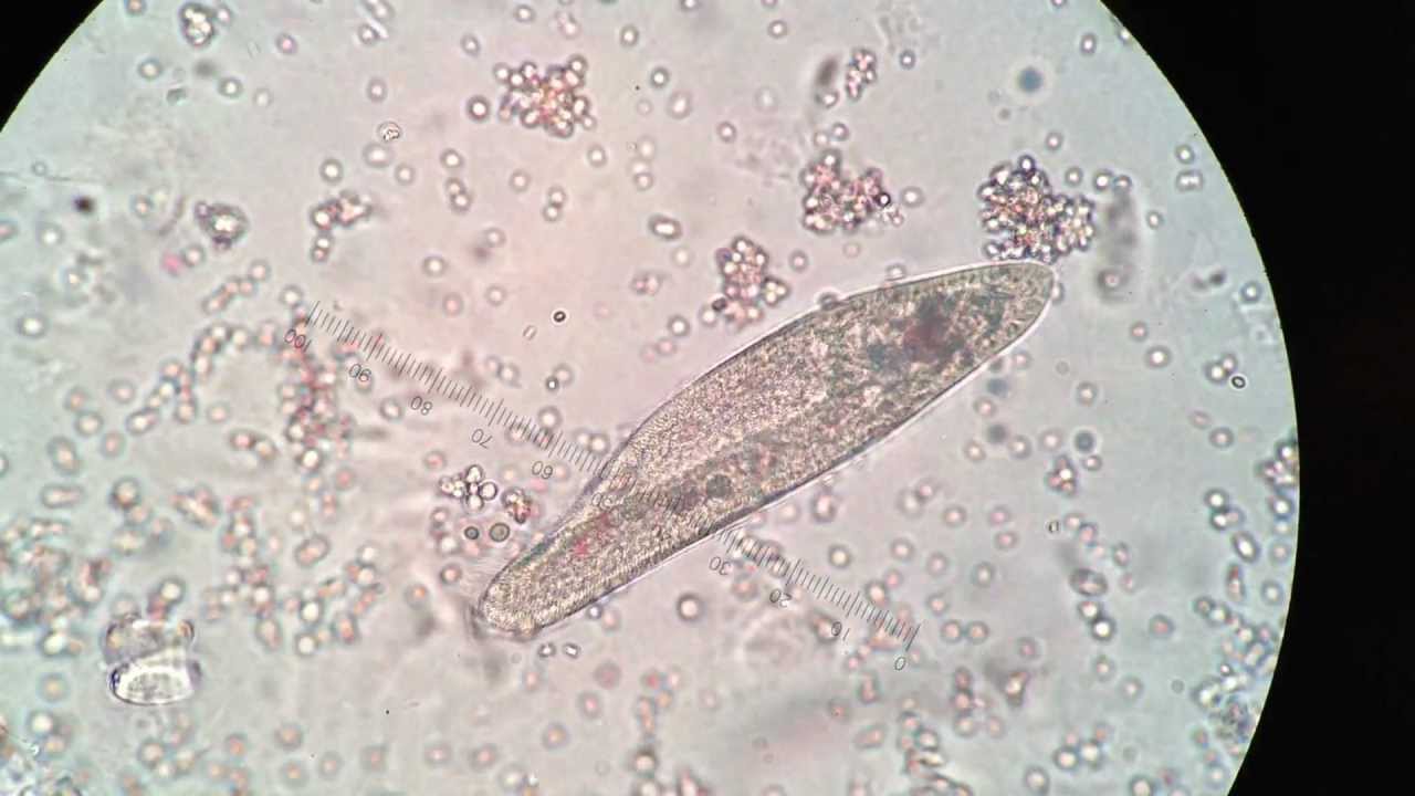
Paramecium under 400X magnification - YouTube
Microscope Labeling Game - PurposeGames.com About this Quiz. This is an online quiz called Microscope Labeling Game. There is a printable worksheet available for download here so you can take the quiz with pen and paper. This quiz has tags. Click on the tags below to find other quizzes on the same subject. Science.

BIO 156, Fall 2015: Week 8. Lab 7. Immune System
Looking at the Structure of Cells in the Microscope The Light Microscope Can Resolve Details 0.2 μm Apart. In general, a given type of radiation cannot be used to probe structural details much smaller than its own wavelength. This is a fundamental limitation of all microscopes. ... and fluorescent labels are usually used for the most precise optical localization.
Nanogold-Antibody Conjugates
Compound Microscope Parts, Functions, and Labeled Diagram Compound Microscope Definitions for Labels. Eyepiece (ocular lens) with or without Pointer: The part that is looked through at the top of the compound microscope. Eyepieces typically have a magnification between 5x & 30x. Monocular or Binocular Head: Structural support that holds & connects the eyepieces to the objective lenses.

Bone Lab
Microscope Parts and Functions Microscope Parts and Functions With Labeled Diagram and Functions How does a Compound Microscope Work?. Before exploring microscope parts and functions, you should probably understand that the compound light microscope is more complicated than just a microscope with more than one lens.. First, the purpose of a microscope is to magnify a small object or to magnify the fine details of a larger ...
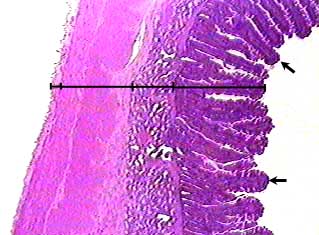
Duodenum
Animal Cell Under Light Microscope Labelled : Draw and label the ... A cell is a very tiny structure which exists in living bodies. .for viewing under the light microscope can label plant and animal cell structures and describe their functions to be able to work out the size of a cell contain chlorophyll which absorb light energy to make food by photosynthesis lipid membrane which controls what enters and leaves ...
302 Found
› news › laboratory-productsThe Importance of Labels in Laboratory Testing Labmate Online May 18, 2022 · Customising your labels. Cryogenic labels need to resist temperatures as low as -196°C. Additionally, the repeated freeze and thaw cycles combined with other harsh processes will at the very least put your label's solvent compatibility to the test. Nitrogen liquid and vapour can be a hazard for labels also.
Post a Comment for "43 light microscope with labels"